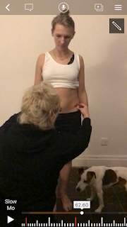
Static Posture Assessment
Static Posture
Postural assessment is used from ages, were a critical element of any
evolution. In current time, when knowledge is developed and evidence-based
medicine progressed It is time to establish if the postural assessment is
effective, however, there are limited clinical studies and evidence-based
whether the postural assessment is effective. Posture assessment can be static
and dynamic Static posture assessment is the view of physical looks in the
mirror, when a person is in stance. Is need of a strong visual observation skill
from the person who does it. Starts from feet and goes upwards towards the head. The
static postural assessment has been the basis to recognize muscle imbalance. (Clark et al., 2014).
Posterior View
is very
similar to the anterior view. This what showed a posterior view, an anterior
view will confirm. The head (ear level), shoulder position, knee interspace,
and ankle position are all assessed in addition to the overall symmetry of the
body (Clark et al., 2014).
The posterior view has given extra information towards a heel position
and scoliosis screening of the spine. Scoliosis is a lateral and from time to
time rotatory curvature of the spine resulting in a "C" or
"S" shaped curve the ankle allows assessment of the heel (Gies et al., 2018).
Plumb line
left side
right side
Client A,
is a 35-year-old female. Mother of two boys aged an 11 and an 8. She works in a
bank from 12 years, five days in week, average of thirty hours. Her work is sedentary and she drives a car every day, everywhere. She does exercise
from many years, it is depended on how often, but she is active. She does
yoga one times a week, zumba twice and she walks with her dog every day at
least thirty minutes. Almost every Sunday she goes with her family for a trip.
She does not complain about low back pain at the moment, but every time when
she stops exercise, low back pain comes back, she is aware that she must exercise
to help herself and she is not getting younger, age is a factor which can make her lower back pain worse.
Head and
neck
Person spins
three times and then stands still. An imaginary line goes from midway between the heels,
extending upward between the lower extremities, through the midline of the
pelvis, through the spine and skull. Head has very small flexion. Ears level is almost the same on both sides. The right side is
slightly lower than the left side. This can point at lateral flexion; the cause
can be shortened muscles on the side the neck is flexed to. The head and neck
tilt looks correct, asymmetrically. There is no cervical rotation and cervical
vertebrae is correct.
Shoulder
height
A person
is a right hand, it is a dominate side where hypotrophy is noticed. Can be that there is tightness in the upper trapezius and levator scapulae muscles on the side, or because is right
hand.
Scapula
Adducted
scapula on the left side, is too close to the midline of the thoracic vertebrae
(only two fingers). The right side: scapula is far from the
midline on three fingers so it is correct distance.
The
position of the inferior angle of the scapula is slightly different. The left
scapula is lower campers to the right which is elevated. Muscles which are responsibility
for elevater the scapula are shorter on the side that elevates the scapula -
upper fibres of the trapezius and lavator scapulae.
Rotation
of the scapula in this situation is a downward rotation. In downwardly rotated
scapulae it will be abducted at the superior angle and adducted at the inferior
angle. The left scapula is bigger and wider. There is no winging of the
scapulae.
Trunk
Lateral
deviation (Scoliosis). The spinous processes of the vertebrae are lateral to
the midline of the trunk. Intrinsic trunk muscles are shortened on one side and
contralateral intrinsic trunk muscles lengthened and compression of the vertebrae
on the concave side. Leg-length discrepancy.
Thoracic
Cage is not
rotate in relation to the client's head and hips.
Upper
Limb Position
Space is
not the same between the client's arms and their body. On the right side there is no
space. The hip hitched and their pelvis is laterally tilted upwards.
Elbow position are almost correct as
well as hands. Palms are facing their thighs, they are not rotated
forward.
Pelvis
and Hip
Lateral pelvic tilt, right side of the pelvis is higher posterior superior iliac. What can point to scoliosis with ipsilateral lumbar convexity, leg-length discrepancies. Can be tight ipsilateral hip abductor muscles on the same side as well as tight contralateral hip adductor muscles and can be a weakness of the contralateral abductor muscles. Lumbar spine - pelvis raised on the right. Flex to the right and concave on the right. In the lumbar muscles can be shortened, right quadratus and lumbar erector spine, this can have an effect on the hip joint which means that the right one is adducted and the left one is abducted. Right hip can be shorter adductors and the left hip opposite abductors, this can point that there is an imbalance between the right and left hamstring. Posterior Iliac Spine (PSIS) is not on the same level which suggests that the client has lateral tilt of the pelvis.
Calf
we can see that the feet are slightly flat going interior - calcaneovalgus (everted-pronated). Weak muscles can be supinators of the foot.
we can see that the feet are slightly flat going interior - calcaneovalgus (everted-pronated). Weak muscles can be supinators of the foot.
Anterior View will confirm posterior view
Plumb line
The head
is deviating slightly, laterally from the midline, is shorter stronger upper
Trapezius, shoulders are not on the same level, one is elevated. Shoulders are
not rounded. The anterior iliac spines (ASIS) is slightly lower than the other
(lateral pelvis). Legs have got different lengths. I took a measurement and one is
longer by 1cm, than the other one and the patella is rotated inwards, it should point straight with the respect to the tibiofemoral joint (Edgerton et al., 1996). The rotation of the patella can be link with rotation of the femur or torsion of the tibia (Vesci et al., 2007). The shorter leg will therefore have to over compensate to work as effectively as the other. Ankles slightly inwards towards the midline of the body - forefoot valgus feet flat (plantarflexed position.)
Abducted hip -hip shift
Client
has hip shift - she has weak hip abductor with compensation reason can be a weakness of contralateral adductors and ipsilateral abductors (Clark et al., 2014).
Lateral View
Plumb line
Client's
head is pushed forward with the chin out from the midline and increased -
lumbar spine, lordotic curve. An anteriorly tilted, it is very often that
during pregnancy time, baby weight which is growing cause the abdominal muscles
to lengthen and weaken, hip extensors, thereby pelvis can change position which can cause be a hyperlordotic posture (Connes et al., 2010).
Client was twice pregnant, she gave natural birth.
It is a
big difference between the height of PSIS and ASIS- which is much lower, pelvic
tilt, but sometimes can be an anatomic structure (Clark et al., 2014).
Knee is
hyperextended - genu Recurvatum, the gravitational stresses lie forward of the
joint axis which can be caused from the tightness of the quadriceps, gastrocnemius, soleus muscle, stretched popliteus and hamstring muscles, at the knee can sometimes
compression forces anteriorly or shape of tibial plateau.
Feet have
arch to low -(pes planus).
After a prolonged amount of weight bearing exercise the client could experience symptoms of plantar fasciitis, 1stMPTJ pain, Metatarsalgia, arch pain and generalised inflammation around soft tissue structures(Vesci et al., 2007).
After a prolonged amount of weight bearing exercise the client could experience symptoms of plantar fasciitis, 1stMPTJ pain, Metatarsalgia, arch pain and generalised inflammation around soft tissue structures(Vesci et al., 2007).
Reference
Connes, P., Hue, O. and Perrey, S. (2010). Exercise physiology. Amsterdam: IOS Press.
Gies, T. (2018). The ScienceDirect accessibility journey: A case study. Learned Publishing, 31(1), pp.69-76.
Sloot, L., van der Krogt, M. and Harlaar, J. (2014). Self-paced versus fixed speed treadmill walking. Gait & Posture, 39(1), pp.478-484.
Micheal A. Clark, Scott C. Lucett, Brian G. Sutton (2014). Corrective Exercise Training. Knee. 28(5), pp100.107.
Vesci BJ, Padua DA, Bell DR, Strickland LJ, Guskiewicz KM, Hirth CJ. Influence of hip muscles strength, flexibility of hip and ankle musculature, and hip muscle activation on dynamic knee valgus motion during a double - egged squat. J Athl Train. 2007;42:S-83


Komentarze
Prześlij komentarz